


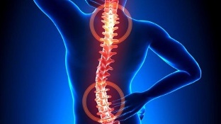
Excessive tension in the back muscles causes a lot of discomfort and pain. Osteochondrosis, which disrupts the structure of the vertebrae and intervertebral discs, causes severe compression of the nerve endings. It is often accompanied by pathological deterioration of blood circulation, which leads to disturbances in the nutrition of the brain and internal organs.
Osteochondrosis - what is it?

Osteochondrosis is a recurrent form of the disease that is chronic and is accompanied by destruction of the vertebrae by intervertebral discs. Their tissues become disturbed, which leads to a decrease in the degree of elasticity, and then a change in shape. There is a gradual decrease in the intervertebral space. This leads to a loss of stability of the spine in areas of pathology.
The process of destruction of pathological tissues occurs against the background of compressed nerve endings directed from the area where the spinal cord is located. As a result, the back muscles are under constant tension. In this case, patients complain of back pain and other symptoms.
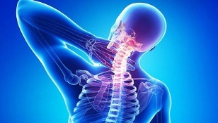
Cervical, thoracic and lumbosacral types of pathological process differ according to the localization features of the spinal structures surrounded by degenerative changes. The main symptom of the development of osteochondrosis is pain, which usually increases in intensity and severity during physical exertion.
There is also rigidity in movement. In addition, the clinical picture is characterized by the presence of vertebral-type symptoms - headaches, changes in blood pressure, impaired vision, hearing, etc.
Development Mechanism
The development of osteochondrosis is associated with the loss of hydrophilic qualities of the pulposus nucleus. This semi-liquid structure contains chondroitin, a substance with connective tissue fibers and gelatin. In the process of development and growth of the human body, the process of reducing the vascular bed in the intervertebral discs continues actively. Foods are given in a diffuse manner, which manifests itself in the stabilization of spontaneous concentration. This feature causes difficulties in the complete recovery of damaged or excessively pressed cartilage on the spine.
Pathological abnormalities are more striking due to hormonal background and human nutritional disorders. Cartilage tissue begins to experience a lack of nutrients required for normal development. Therefore, the disorders appear as follows:
- decrease in strength and elasticity;
- Changes consistency parameters and configuration properties.
Radial cracks appear in the annular fibrosis against the background of flattening of the intervertebral discs. As a result, the distance between the vertebrae decreases and the facet joints begin to change. Pathological changes over time include types of connective tissue associated with fibrous rings and ligaments.
As tissues break down by the immune system, increasing amounts of immunoglobulin are formed. This leads to the development of aseptic inflammatory process, edema occurs in the area where the facet joints are located. They also spread to adjacent soft tissues.
Due to the elongation of the connecting capsules, the intervertebral discs lose their ability to straighten the spine. This instability of the spinal structure increases the risk of squeezing nerve roots or squeezing blood vessels. This feature is typical, for example, of cervical osteochondrosis, which is accompanied by intense oral symptoms.
Causes of the disease
The condition of the intervertebral discs may worsen with a decrease in the tone of the muscles of the spine. Due to the irrational and asymmetrical work of the muscles, the destruction of cartilage tissue can occur with long-term protection of the non-physiological state of the body. This violation is the result of wearing heavy bags on the same shoulder, using soft mattresses and high pillows.
The process of destruction of intervertebral discs is accelerated by a number of negative factors of external and internal nature. These include:
- disorders of the endocrine mechanism and metabolic diseases;
- a pathology of an infectious nature, including a chronic form;
- compression fractures of the spine, bruises;
- regular and long-term hypothermia of the body;
- systemic and degenerative-dystrophic type diseases - gout, psoriatic, rheumatoid arthritis, osteoporosis, osteoarthritis;
- smoking and alcohol abuse, which impairs the condition of the vascular system, worsens blood circulation and causes malnutrition in cartilage;
- Insufficient physical development, posture problems, flat feet - these defects increase the load on the spine, because the cushioning will not be enough;
- obesity;
- genetic predisposition;
- regular stress.
symptoms

The main clinical sign of osteochondrosis of any localization (cervical, thoracic or lumbosacral) is pain syndrome. With a recurrence, the pain penetrates and spreads to nearby areas of the body. It intensifies even with a small action. This forces the patient to force the body to minimize discomfort and pain:
- With cervical osteochondrosis, it is preferable to turn the whole body, not just one head;
- When the disease is in the thoracic form, it is difficult for the patient to breathe deeply, and therefore tries to minimize the depth and frequency of breathing to exclude acute pain in the chest;
- Patients with low back pain have difficulty sitting, standing, or moving because the nerves in the spine are constricted.
Typically, patients complain of motionless, persistent pain and stiffness in the morning after waking up. In this case, a differential diagnosis will be required to help rule out the risk of developing myositis resulting from inflammation of the skeletal muscles or osteoarthritis.
Pain and cramping pain are caused by compensatory tension in the muscle tissues. This position is necessary to stabilize the area of spinal movement. Persistent mild to moderate pain may occur with significant elongation of the intervertebral disc and may result in aseptic inflammatory changes.
Osteochondrosis of a separate localization is characterized by specific symptoms:
- Pain with cervical osteochondrosis is felt in the cervical region, in the upper extremities. Headaches and numbness of the fingers are observed. If the disease is severe, the vertebral artery may constrict. In this case, the patient begins to complain of significant deterioration of health.
- Thoracic localization, manifested by sharp and painful pain in the back, visceral pain syndrome in the heart, right hypochondrium and abdomen. Patients complain of numbness, paresthesia of the skin, shortness of breath, tightness in the vertebrae.
- Patients with lumbar osteochondrosis complain of increased pain in the lower back and lower extremities when moving. Often, disorders of the genitourinary system, problems with male potential, non-functional ovarian disorders are diagnosed. Pain may be reduced during remission. However, the effect of a provocative factor causes its renewal.
- When mixed osteochondrosis manifests itself, the symptomatology may manifest itself in several zones at the same time. This condition is characterized by a more severe course of the disease.
Recall that the displacement of the vertebrae and the formation of osteophytes cause compression of the vertebral artery. It nourishes the brain, which provides its cells with an oxygen component. When squeezed, food is limited, and therefore the patient has problems with coordination, headache, tinnitus and arterial hypertension.
Untreated outcomes
The reason for the complex course of osteochondrosis is the relatively rapid formation of hernias in the intervertebral discs. Their appearance is associated with the posterior displacement of the vertebral structure. This leads to a rupture of the longitudinal posterior ligament, resulting in instability of the position of the disc, resulting in the protrusion of individual parts in the region of the spinal canal. Hernia rupture occurs when a nucleus with a pulposus penetrates the canal area.
With the manifestation of pathological abnormalities in the vertebral structures, the back of the brain begins to constrict, the patient develops discogenic myelopathy. Symptoms of this condition are associated with numbness and weakness in certain muscle groups of the upper and lower extremities. Paresis, muscle atrophy and tendon reflexes manifest themselves. In some cases, there are problems with emptying the bladder and intestines.
Herniated discs are dangerous by squeezing the blood vessels that supply the spinal cord. The result of this pathology is the formation of ischemic zones where nerve cells are damaged and killed. Manifestations of neurological effects are characterized by impaired motor function, decreased sensitivity and trophism.
Diagnostics
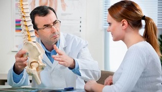
Initial diagnosis is based on the patient's complaints and symptoms. The specialist examines the condition of the spine in different positions and suggests that the patient be at rest or in motion. The next step is referring to laboratory diagnostics that will help the patient confirm or refute the diagnosis.
The research methods used include:
- Radiography- provides a complete examination of the spine, the condition of the vertebrae, with the assessment of existing disorders in the form of growths, curvatures. The specialist will be able to determine the intervertebral type breaks, the condition of the holes. A two-stage X-ray examination is performed to accurately diagnose localized osteochondrosis in the chest or neck. In the first stage, the patient lies on his side, and in the second directly behind.
- MRI or CT tomographyprovides highly informative information that helps to study in detail without interfering with the shape of the organs surrounding the spine. The picture shows the nervous and vascular system. MRI helps to identify the symptoms and location of damage in many diseases of the spine. CT scans show hernias, identify possible deviations in the structure of the spine.
- Laboratory examination to assess the condition and basic parameters of the blood. Allows to clarify the diagnosis and determine the development of concomitant diseases.
In many cases, as a result of examinations, doctors diagnose some background diseases that are potentially dangerous for complications. For example, we are talking about hernias, bulges, radiculitis. Proper diagnosis of problems helps to effectively treat osteochondrosis. At the same time, the disease itself in the early stages of development is hidden as a symptom of other diseases.
Therapeutic Process
Osteochondrosis is treated conservatively or surgically. The choice depends on the severity of the condition, indifference, the degree of tissue deterioration and the reasons.
It is important to remember that osteochondrosis cannot be completely cured because there are no medications to help the discs and spine recover completely. The therapeutic effect is aimed at preventing the process of destruction and increasing the duration and stability of remission.
Chondroprotectors based on chondroitin sulfate or glucosamine are used for symptomatic therapy.
The effectiveness of the therapeutic process using chondroprotectors has been clinically confirmed on the basis of long-term tests. If you take these funds for a long time from 3 months, there is a partial recovery of cartilage and other elements of the connective type - the garden-tendon apparatus, bursa.

Accumulation of glucosamine and chondroitin in the area of the intervertebral discs causes analgesic, anti-edematous and anti-inflammatory effects. Therefore, there is a real opportunity to optimize the dose of NSAIDs, glucocorticosteroid drugs, muscle relaxants. You can hope to reduce the burden of medication on the patient.
The effectiveness of chondroprotectors is determined by the rules of their reception. Otherwise, there will be no result. Efficacy is also observed in the treatment of grade 3 osteochondrosis, which is accompanied by significant destruction of cartilage.
The following groups of drugs can be used to reduce pain:
- Non-steroidal anti-inflammatory drugsHelps relieve inflammatory disorders in soft tissues caused by vertebral displacement. NSAIDs are effective in reducing pain, swelling and stiffness.
- Glucocorticosteroids- blockers are usually used in combination with anesthesia. They can relieve pain, restore the immune mechanism and have an exudative effect.
- Muscle relaxants.They are effective against muscle spasms due to nervous breakdowns. Helps relax skeletal muscles and binds polysynaptic spinal type reflexes with antispasmodic effect.
- An external agent with a warming effect.Irritation of subcutaneous tissue receptors by activation of blood flow is provided by special gels and ointments. These drugs have analgesic and anti-edematous effects.
With the help of medical devices to activate blood flow, it is possible to eliminate the symptoms of vertebrogenic type caused by the localization of the pathology in the cervical or thoracic region. Nootropics and medications are also prescribed to improve microcirculation. In some cases, you may need to take medications that contain antidepressants as well as anticonvulsants.
Physical therapy is also used in the treatment of osteochondrosis. UHF therapy, magnetotherapy, laser therapy, reflexology, massage, sports therapy, hirudotherapy, as well as swimming and yoga procedures can be prescribed. If conservative treatment is not effective, the operation is performed by microdiscectomy, valorization of the perforated disc, laser reconstruction or implant replacement.