


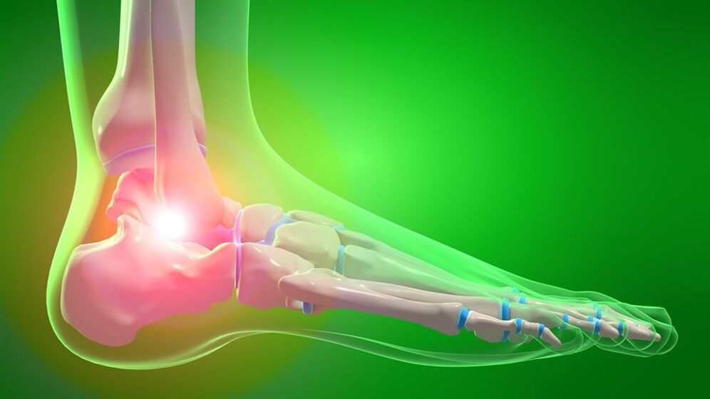
Arthrosis of the ankle joint is a chronic disease that affects the articular cartilage and subsequently other structures of the joint (capsule, synovium, bones, ligaments). It has a degenerative-dystrophic nature. It manifests as pain and limitation of movements, followed by progressive impairment of support and walking functions. Diagnosis is based on symptoms, examination and X-ray. Treatment is usually conservative, anti-inflammatory drugs, chondroprotectors and glucocorticoids are used, exercise therapy and physical therapy are prescribed. In severe cases, sanitary arthroscopy, arthrodesis or endoprosthetics are performed.
General Information
Osteoarthritis of the ankle joint is a disease in which joint cartilage and surrounding tissues are gradually destroyed. The disease is based on degenerative-dystrophic processes, inflammation in the joint is secondary. Arthrosis has a chronic wave-like course with alternating remission and exacerbation and progresses gradually. Women and men suffer equally often. The probability of development increases dramatically with age. At the same time, experts note that the disease is "getting younger" - now every third case of ankle arthrosis is detected in people under 45 years old.
Reasons
Primary arthrosis occurs for no apparent reason. Secondary damage to the ankle joint develops under the influence of some unfavorable factors. In both cases, the main factor is the disruption of metabolic processes in the cartilage tissue. The main causes and predisposing factors of secondary arthrosis of the ankle joint:
- large intra- and periarticular injuries (fractures of the talus, fractures of the ankle, tears and ruptures of ligaments);
- ankle surgery;
- excessive load: very intense sports, long walks or standing constantly due to work conditions;
- wearing shoes with heels, overweight, constant microtraumas;
- diseases and conditions associated with metabolic disorders (diabetes, gout, pseudogoat, estrogen deficiency in postmenopause);
- rheumatic diseases (SLE, rheumatoid arthritis);
- osteochondrosis of the back, intervertebral hernia and compressed nerves, and other conditions accompanied by a violation of the muscular system of the foot and leg.
In less cases, the cause of arthrosis is arthritis due to non-specific purulent arthritis, specific infections (tuberculosis, syphilis) and congenital developmental anomalies. Unfavorable environmental conditions and hereditary predisposition play a certain role in the development of arthrosis.
Pathogenesis
Normally, the articular surfaces are smooth, elastic, slide smoothly relative to each other during movements, and provide effective shock absorption under load. As a result of mechanical damage (trauma) or metabolic disorders, cartilage loses its smoothness, becomes rough and elastic. During movements, the cartilage "rubs" and injures each other, which leads to the worsening of pathological changes.
Due to sufficient wear, excess load is transferred to the main bone structure and degenerative-dystrophic disorders also develop: the bone is deformed and grows along the edges of the articular region. Due to secondary trauma and disruption of the normal biomechanics of the joint, not only the cartilage and bone, but also the surrounding tissues suffer.
The joint capsule and synovial membrane thicken, fibrous degeneration foci are formed in ligaments and periarticular muscles. The joint's ability to participate in movements and withstand loads decreases. Instability develops and pain progresses. In severe cases, articular surfaces are destroyed, the supporting function of the limb is disturbed, movements become impossible.
Symptoms
At the beginning, after a significant load, rapid fatigue and mild pain are detected in the ankle joint. Later, the pain syndrome becomes stronger, its nature and time of occurrence change. Distinctive features of pain with arthrosis are:
- Onset pain. It appears after a state of rest and then gradually disappears with movement.
- Load dependence. There is increased pain during exercise (standing, walking) and rapid fatigue of the joint.
- Night pain. It usually appears in the morning.
The condition changes in waves, during the exacerbation the symptoms are more obvious, in the phase of remission they first disappear, and then become less intense. There is a gradual progression of symptoms over several years or decades. In addition to pain, the following manifestations are identified:
- There may be creaking, creaking, or clicking noises when moving.
- During an exacerbation, the periarticular area sometimes swells and turns red.
- Due to the instability of the joint, the patient often twists the leg, causing sprains and tears in the ligaments.
- Stiffness and limitation of movements are noted.
Complications
During an exacerbation, reactive synovitis may occur, accompanied by fluid accumulation in the joint. In the later stages, a clear deformation is detected. Movements are sharply limited and contractures develop. Support becomes difficult, when moving, patients are forced to use crutches or crutches. There is a decrease or loss of working capacity.
Diagnostics
The diagnosis of arthrosis of the ankle joint is made by an orthopedic doctor based on a questionnaire, external examination data and the results of additional studies. In the initial stages, there may be no changes when examined, but later deformations, restriction of movements, and pain during palpation are revealed. The main importance is given to visualization methods:
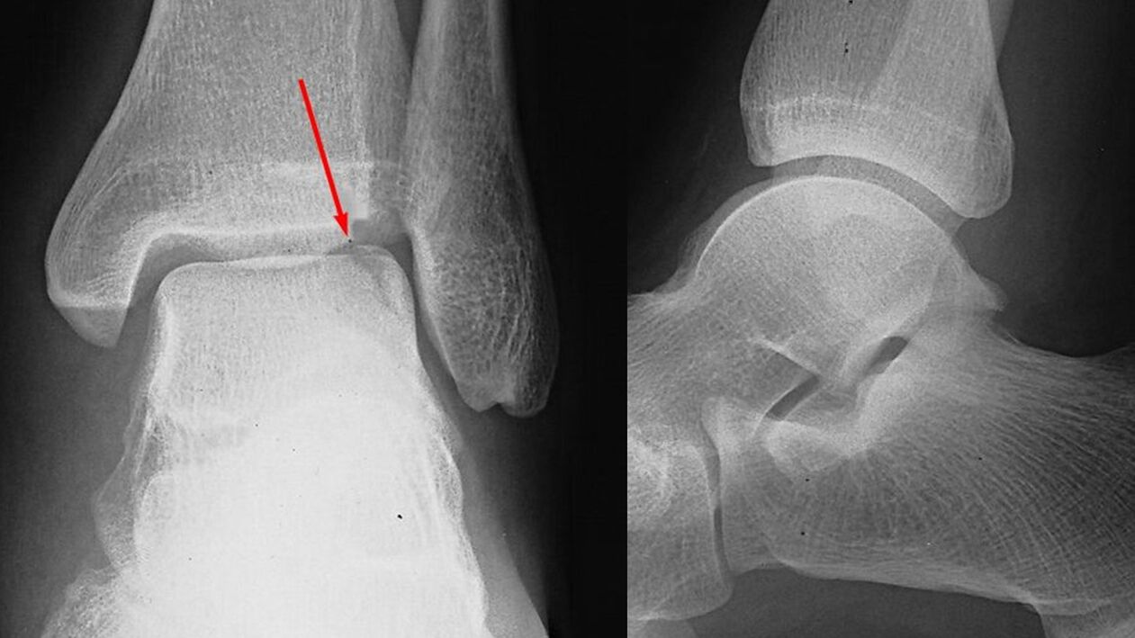
- X-ray of the ankle joint. It plays a decisive role in making a diagnosis and determining the degree of arthrosis. Narrowing of the pathological joint space is indicated by the spread of the edges of the articular surfaces (osteophytes). At a later stage, cystic formations and osteosclerosis of the subchondral (located under the cartilage) zone of the bone are detected.
- Tomographic studies. Used when indicated. In difficult cases, in order to more accurately evaluate the condition of the bone structures, the patient is additionally sent for computed tomography and examination of soft tissues - MRI of the ankle joint.
Laboratory tests are unchanged. If necessary, consultations with related specialists are prescribed to determine the cause of arthrosis and differential diagnosis with other diseases: neurologist, rheumatologist, endocrinologist.
Treatment of ankle arthrosis
Treatment of pathology is long-term and complex. Patients are usually seen by an orthopedic surgeon on an outpatient basis. Hospitalization in the department of traumatology and orthopedics is possible during the period of exacerbation. The most important role in slowing down the development of arthrosis is played by lifestyle and proper physical activity regime, so the patient is advised to lose weight and optimize the load on the foot.
Drug therapy
The stage of arthrosis is selected individually, taking into account the severity of symptoms and accompanying diseases. Includes general and local agents. The following drug groups are used:
- Generic NSAIDs. Tablet forms are usually used. Medicines have a negative effect on the gastric mucosa, so "gentle" medicines are preferred for gastrointestinal diseases.
- Topical NSAIDs. It is recommended both in the period of exacerbation and in the phase of remission. If side effects occur from tablet forms, they can be prescribed as an alternative. It is available in the form of ointment and gel.
- Chondroprotectors. Substances that help normalize metabolic processes in cartilage tissue. They are used in the form of creams, gels and preparations for intra-articular administration. Use medications containing glucosamine and collagen hydrolyzate.
- Hormonal agents. In cases of severe pain that cannot be relieved by drugs, intra-articular corticosteroids are administered no more than 4 times a year.
- Metabolic stimulants. Nicotinic acid is prescribed to improve local blood circulation and activate tissue metabolism.
Physiotherapy treatment
The patient is prescribed a complex of physical therapy designed taking into account the manifestations and stage of the disease. The patient is sent to physiotherapy. Massage and UHF are used in the treatment of arthrosis. In addition, they are used in the treatment of pathology:
- laser therapy;
- thermal procedures;
- drug electrophoresis and ultraphonophoresis.
Surgery
When conservative therapy is ineffective, severe pain syndrome, deterioration of patients' quality of life or limitation of work capacity are indicated in the later stages of the disease. Operations are performed in a hospital setting and are open and minimally invasive:
- Arthroscopic interventions. Arthroscopic chondroplasty is performed in case of significant cartilage destruction. Sanitary arthroscopy (removal of formations that interfere with movement) is usually performed for severe pain in the 2nd stage of arthrosis. Its effect lasts for several years.
- Arthrodesis of the ankle joint. It is carried out in case of significant destruction of the articular surfaces, involves the removal of the joint and the "fusion" of the bones of the leg and lower leg. It ensures the restoration of the supporting function of the limb during the loss of joint mobility.
- Endoprosthesis of the ankle joint. It is performed for advanced arthrosis. It involves removing the destroyed articular surfaces of the bones and replacing them with plastic, ceramic or metal prostheses. Movements are fully restored, the service life of the prosthesis is 20-25 years.
Forecast
The changes in the joint are irreversible, but the slow progression of arthrosis, the timely start of treatment and following the recommendations of an orthopedic traumatologist in most cases allow to maintain working capacity and a high quality of life for decades after the appearance. from the first symptoms. With the rapid increase in pathological changes, endoprosthetics allow to avoid disability.
Prevention
Preventive measures include reducing the level of injuries, especially in winter, during the ice age. If you are obese, you should take measures to reduce your body weight to reduce the burden on your joints. You should maintain a moderate physical activity regime, avoid overloads and microtraumas, and immediately treat diseases that can lead to the development of arthrosis of the ankle joint.